Or call us at 209-879-9367 — we'll answer Mon–Fri, 8 AM to 4:30 PM.📍 1973 N. Tracy Blvd., Tracy, CA 95376
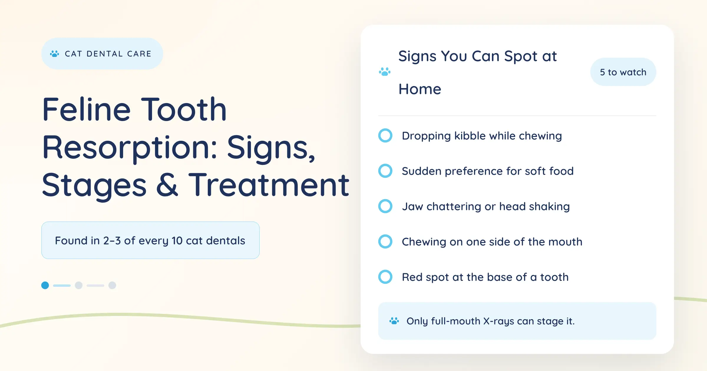
Schedule OnlineYou notice your cat dropping kibble she used to crunch without hesitation, or you catch a small red spot low on a tooth near the gum line. These are two of the earliest owner-visible signals of feline tooth resorption.
At The Pet Advocate we see this condition regularly. When we perform 10 cat dental procedures, we typically find resorptive lesions in two to three of those patients. The process is progressive. Here is what feline tooth resorption actually is, the signs you can check at home, how we diagnose and treat it, and what to expect at our Tracy clinic.
Written and reviewed by Michael Wesselink, DVM – August 2026
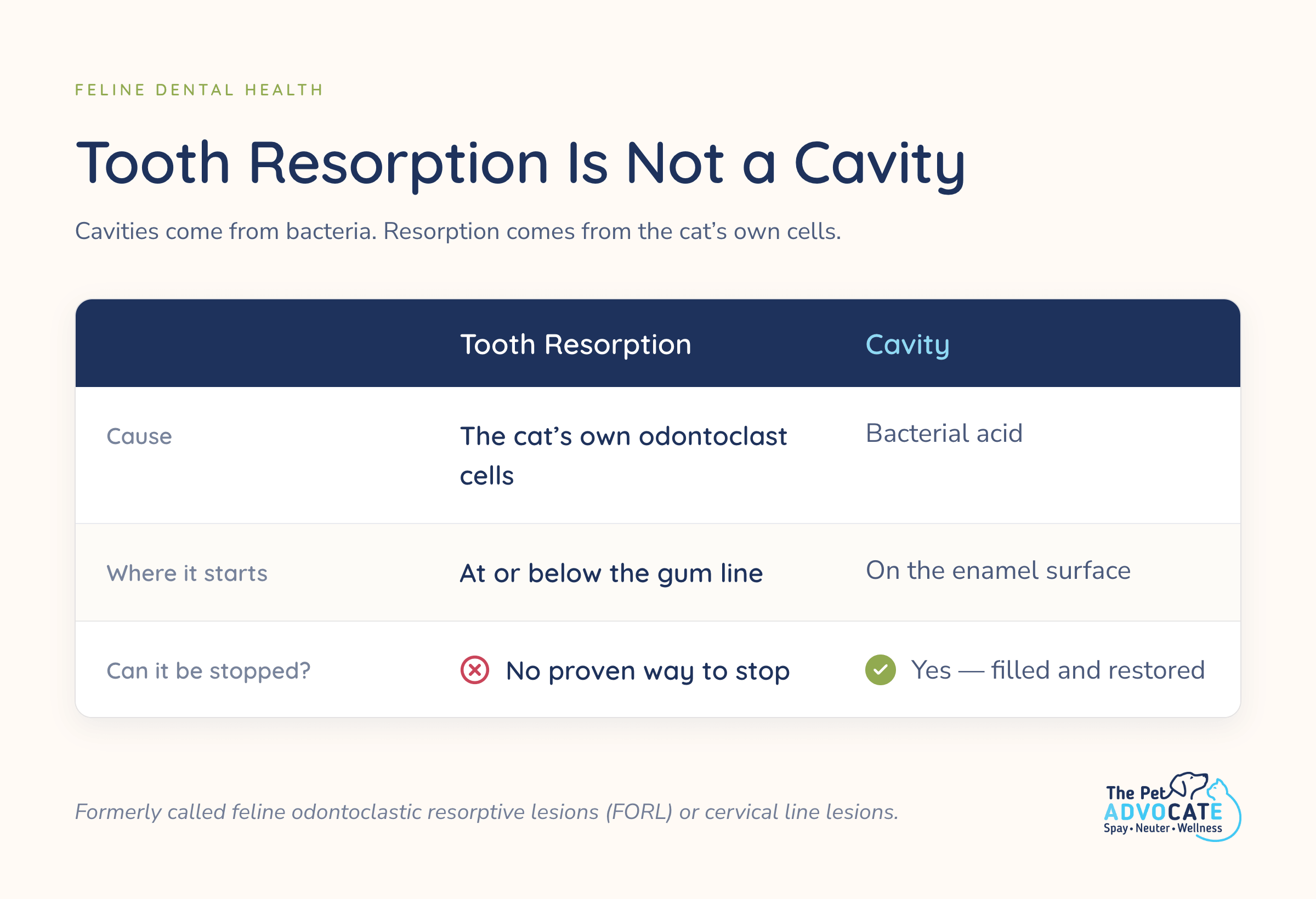
Feline tooth resorption is an inflammatory process in which the body begins breaking down the hard structures of a tooth. Odontoclast cells, which normally help shed baby teeth, attack the cementum, dentin, and eventually the crown or roots of permanent teeth.
The result is holes or lesions that start below or at the gum line and progress. The condition was previously called feline odontoclastic resorptive lesions (FORL) or cervical line lesions. It is almost exclusively a cat problem and is very rare in dogs. Owners of both species can still protect oral health with routine dog dental cleaning and cat dental care.
Feline tooth resorption is not a cavity. Cavities are caused by bacterial acid. Resorption is driven by the cat’s own cells. There is currently no proven way to stop the process once it starts.
Population studies place lifetime prevalence between roughly 20% and 75% of cats, with risk rising sharply after age five. The Cornell Feline Health Center notes that close to three-quarters of cats five years and older may be affected. Purebred cats may show lesions earlier, but any cat can develop them.
At The Pet Advocate the practical number is clearer. Two to three of every ten cats that come in for a dental cleaning already have visible or radiographic resorptive lesions. Once one tooth is affected, additional teeth often develop lesions over time.
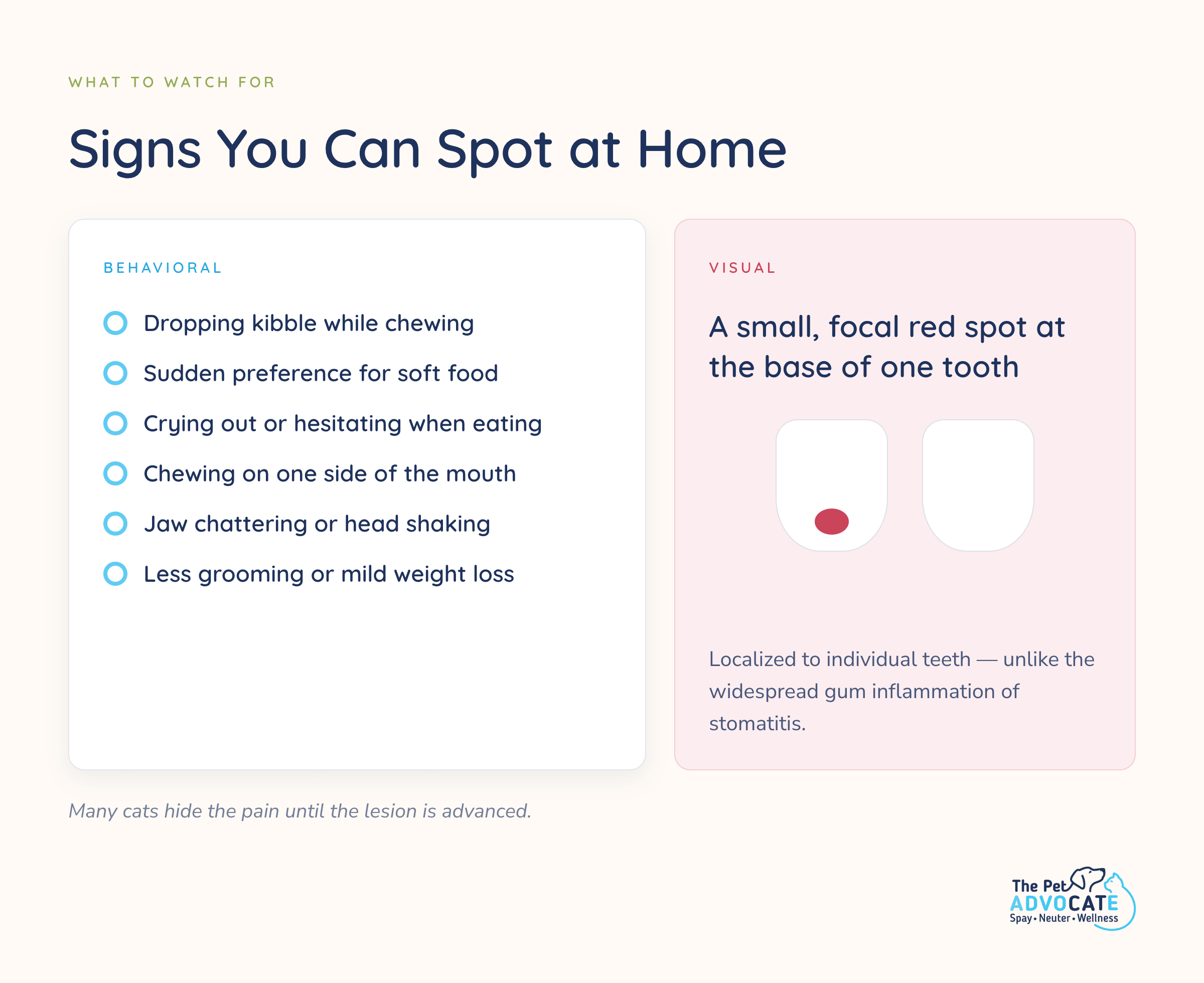
Many cats hide the pain of feline tooth resorption until the lesion is advanced. The most useful early signals are behavioral and visual.
Behavioral signs include:
Visual signs appear as small, focal red spots on the tooth surface, most often at the base near the gum line. These red spots are different from the diffuse, widespread gum inflammation seen in stomatitis. The spots are localized to individual teeth.
If you lift the lip and see a red lesion at the cementoenamel junction or notice your cat changing how she eats, schedule a professional dental evaluation that includes full-mouth radiographs. This presentation is also different from the reversible red-gum inflammation of simple gingivitis, which responds to plaque control when caught early.
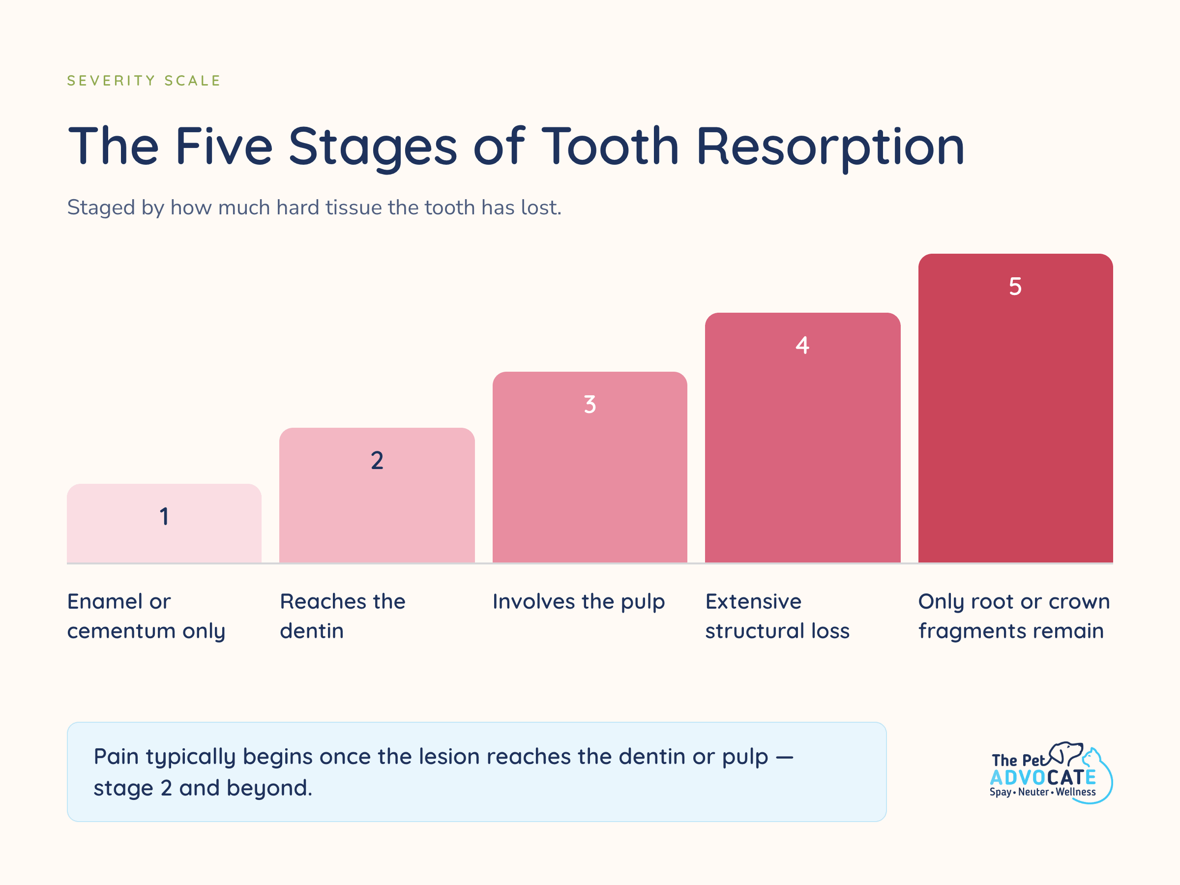
Veterinary dentists classify feline tooth resorption lesions by how much hard tissue is lost (stages 1–5) and by radiographic appearance (Type 1, Type 2, or mixed).
Stage 1 lesions are small and limited to enamel or cementum. Stage 2 reaches the dentin. Stage 3 involves the pulp. Stage 4 shows extensive structural loss. Stage 5 leaves only residual root or crown fragments.
Radiographic type determines treatment:
Full-mouth digital radiographs under anesthesia are required to classify the lesion correctly and plan treatment. Visual inspection alone underestimates the extent of disease. These classifications follow American Veterinary Dental College guidelines and align with the clinical approach outlined in the Merck Veterinary Manual.
Diagnosis of feline tooth resorption starts with an awake oral exam, but definitive classification requires anesthesia, probing, charting, and full-mouth X-rays. Visual inspection alone underestimates how far the lesions have progressed.
At The Pet Advocate the process follows our standard cat dental cleaning workflow. Clients submit an online history form plus three clear photos of the teeth (front and both sides). Those photos are used for initial triage. A $50 deposit holds the appointment.
On the day of the procedure the cat arrives at 8 AM after fasting from midnight. An RVT gathers car-side history. The veterinarian performs a pre-anesthetic physical and preliminary oral exam with the owner present. In-house bloodwork is reviewed and typically takes 15 to 20 minutes.
Once the cat is under anesthesia, the technician charts and probes every tooth. Full-mouth digital radiographs are taken. The veterinarian reviews the images. If extractions are indicated the owner is called for consent before proceeding. This sequence ensures a tooth is never removed without clear radiographic justification and owner agreement. Pre- and post-procedure photos are provided so the difference is visible. You can book an appointment online when you are ready.
Feline tooth resorption has no medical cure that preserves the tooth. The only effective treatment is removal of the painful or exposed portion of the affected tooth.
Lesions are classified radiographically as Type 1 or Type 2. In Type 1 lesions the periodontal ligament remains visible and the root structure is largely intact, so the entire tooth (crown and all roots) must be surgically extracted. Nerve blocks are placed so the cat remains comfortable. The sites are sutured. Post-extraction radiographs confirm complete removal.
In carefully selected Type 2 lesions (advanced root replacement by bone-like tissue, no concurrent periodontal disease, endodontic disease, or stomatitis), a crown amputation below the gum line with intentional root retention and gingival closure may be performed under American Veterinary Dental College criteria. Restoration or filling is not recommended. Filling does not stop the resorptive process and can leave residual pain.
Left untreated, the lesions of feline tooth resorption continue to progress, the cat remains in pain, and additional teeth frequently become involved. After extraction or crown amputation the cat recovers under monitoring and is typically ready for pickup about two hours after the procedure ends. Pain medication is sent home when extractions are performed. Because the disease is progressive, periodic recheck examinations and radiographs are recommended for any cat that has already had one or more lesions treated. Current prices and rates for cleanings and extractions are listed on our site.
There is currently no proven method to prevent feline tooth resorption. Daily brushing and VOHC-accepted dental products reduce plaque and gingivitis, but they do not stop the odontoclast process that drives resorption.
The practical strategy is early detection. Annual professional oral exams and full-mouth radiographs when any suspicious lesion or behavioral change appears allow treatment before the cat experiences prolonged pain. Cats that have already had one resorptive lesion need closer monitoring because additional teeth frequently become affected. At The Pet Advocate our cat dental cleaning process includes the radiographs needed for this monitoring, and you can review current options on our prices and rates page.
Research into the underlying trigger is ongoing. Until a preventive option exists, extraction remains the reliable way to eliminate the source of pain.
Any cat showing dropping of food, a new soft-food preference, jaw chattering, or a visible red spot at the base of a tooth should be evaluated for feline tooth resorption. Cats five years and older benefit from routine professional dental care even without obvious signs, because many lesions are hidden below the gum line until radiographs are taken.
At The Pet Advocate our cat dental cleaning includes pre-anesthetic bloodwork, full-mouth digital X-rays, ultrasonic scaling, polishing, fluoride, charting, and the oral exam. The base cleaning fee is $700. When bundled with a spay or neuter the dental portion is $650. Extractions, when needed, are discussed with you during the procedure and priced accordingly. Most cases that require extractions fall in a predictable mid-range all-in cost of roughly $800 to $1,200. Full details appear on our prices and rates page.
You can book online or by phone. We serve Tracy and the surrounding Central Valley and East Bay communities. See our full list of locations we serve.
Feline tooth resorption is a progressive condition in which the cat’s own cells break down the hard tissues of one or more teeth, creating painful lesions that start at or below the gum line. It is not caused by bacteria the way cavities are.
Yes. Once the lesion reaches the dentin or pulp the tooth is painful, even if the cat continues to eat. Many cats hide the discomfort until the lesion is advanced, which is why dropping food or a red spot at the gum line are important early signals.
No. The process cannot be reversed and fillings or restorations are not recommended. The only effective treatment is extraction (or, in carefully selected Type 2 cases, crown amputation) of the affected tooth.
Population estimates range from 20% to 75% of cats over a lifetime, with higher rates after age five. At The Pet Advocate we see lesions in roughly two to three of every ten cats that come in for a professional cleaning.
Type 1 shows a normal periodontal ligament on radiographs and requires complete extraction of the tooth and roots. Type 2 shows replacement of the root by bone-like tissue; under strict criteria a crown amputation may be appropriate. Full-mouth X-rays are required to distinguish the two.
Brushing and plaque control help with gingivitis and periodontal disease but do not prevent the odontoclast-driven resorption process. Early professional detection with radiographs remains the most useful strategy.